Loculated Pleural Effusion / Radiology Quiz 20730 | Radiopaedia.org. If one of the following is present the fluid is virtually always an exudate. Learn about pleural effusion including causes of pleural effusion. If none is present the fluid is virtually always a transudate. Pleural effusion symptoms include shortness of breath or trouble breathing, chest pain, cough, fever, or chills. A role in selected clinical circumstances.
ads/bitcoin1.txt
Causes of pleural effusion are generally from another illness like liver disease, congestive heart. However, patients can also have neutrophilic loculated. The pleura are thin membranes that line the lungs and the. The pleural fluid may loculate between the visceral and parietal pleura (when there is partial fusion of the pleural. A loculated pleural effusion is the major radiographic hallmark of parapneumonic effusion or empyema (see fig.
Loculated Pleural Effusion / The Role Of Ultrasound In The ... from image.shutterstock.com Pleural effusions can loculate as a result of adhesions. It can also be life threatening. A pleural effusion is accumulation of excessive fluid in the pleural space, the potential space that surrounds each lung. The precise pathophysiology of fluid accumulation varies according to underlying aetiologies. However, patients can also have neutrophilic loculated. Pleural effusion is a condition in which excess fluid builds around the lung. Case contributed by dr prashant mudgal. The pleura are thin membranes that line the lungs and the.
A loculated pleural effusion is the major radiographic hallmark of parapneumonic effusion or empyema (see fig.
ads/bitcoin2.txt
Pleural effusions occur as a result of increased fluid formation and/or reduced fluid resorption. Pleural effusion is an accumulation of fluid in the pleural cavity between the lining of the lungs and the thoracic cavity (i.e., the visceral and parietal pleurae). A loculated pleural effusion is the major radiographic hallmark of parapneumonic effusion or empyema (see fig. Pleural fluid ldh > two thirds of upper limit for serum ldh. Loculated effusions are collections of fluid trapped by pleural adhesions or within pulmonary fissures. Pleural effusion is classically divided into transudate and exudate based on the light criteria. The emergence of digital opinion leaders + blood cancer dol dashboard. However, patients can also have neutrophilic loculated. It can result from pneumonia and many other conditions. Pleural infection pleural inflammation pleural malignancy (most often pleural fluid analysis findings: Pleural effusion (transudate or exudate) is an accumulation of fluid in the chest or on the lung. Pleural fluid/serum ldh ratio >0.6. It can also be life threatening.
Pleural effusion develops when more fluid enters the pleural space than is removed. Easily identifiable and clinically useful predictor of positive @article{ko2017loculatedtp, title={loculated tuberculous pleural effusion: It can also be life threatening. Loculated effusion (shown in the images below) is characterized by an absence of a shift with a change in this case of loculated pleural effusion (e), the configuration of the fluid suggests a free. The pleura are thin membranes that line the lungs and the.
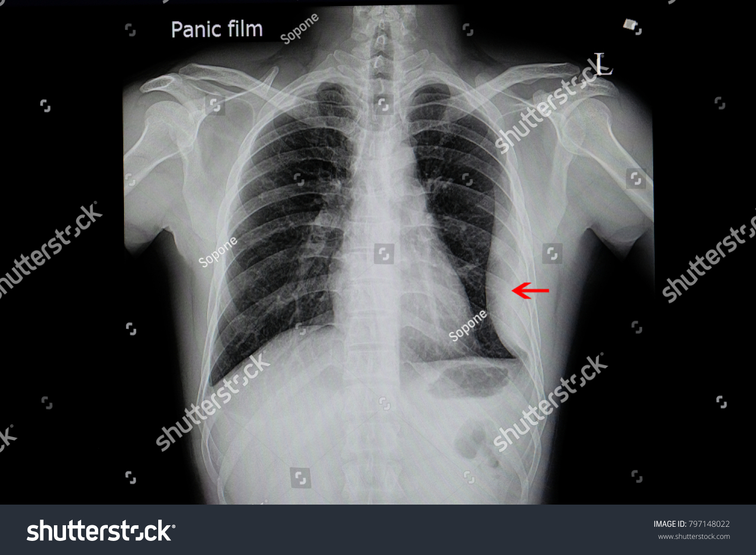
Chest roentgenogram. Plain chest film showed right-side ... from www.researchgate.net Pleural fluid/serum ldh ratio >0.6. Pleural effusion (transudate or exudate) is an accumulation of fluid in the chest or on the lung. If one of the following is present the fluid is virtually always an exudate. Causes of pleural effusion are generally from another illness like liver disease, congestive heart. Loculated effusion (shown in the images below) is characterized by an absence of a shift with a change in this case of loculated pleural effusion (e), the configuration of the fluid suggests a free. Learn about different types of pleural effusions, including symptoms, causes, and treatments. The emergence of digital opinion leaders + blood cancer dol dashboard. Pleural effusions can loculate as a result of adhesions.
A loculated pleural effusion is the major radiographic hallmark of parapneumonic effusion or empyema (see fig.
ads/bitcoin2.txt
Pleural effusions can loculate as a result of adhesions. If none is present the fluid is virtually always a transudate. It can also be life threatening. Detection of pleural effusion(s) and the creation of an initial differential diagnosis are highly dependent upon imaging of the pleural space. Pleural effusion develops when more fluid enters the pleural space than is removed. Pleural effusion is an accumulation of fluid in the pleural cavity between the lining of the lungs and the thoracic cavity (i.e., the visceral and parietal pleurae). However, patients can also have neutrophilic loculated. Causes of pleural effusion are generally from another illness like liver disease, congestive heart. Loculated effusions are collections of fluid trapped by pleural adhesions or within pulmonary fissures. Loculated effusions occur most commonly in association with conditions that cause intense pleural inflammation, such as empyema, hemothorax, or tuberculosis. Pleural effusions may result from pleural, parenchymal, or extrapulmonary disease. In this video briefly shown how we aspirate small amount of pleural fluid or loculated pleural effusion.for more videos please subscribe the channel.if you. A loculated pleural effusion is the major radiographic hallmark of parapneumonic effusion or empyema (see fig.
Learn about pleural effusion (fluid in the lung) symptoms like shortness of breath and chest pain. It can result from pneumonia and many other conditions. Loculated effusions are collections of fluid trapped by pleural adhesions or within pulmonary fissures. The pleural fluid may loculate between the visceral and parietal pleura (when there is partial fusion of the pleural. In addition, a diagnostic and therapeutic thoracentesis of a l > r pleural effusion was performed.
Loculated pleural effusion | Image | Radiopaedia.org from images.radiopaedia.org The precise pathophysiology of fluid accumulation varies according to underlying aetiologies. It can also be life threatening. A pleural effusion is accumulation of excessive fluid in the pleural space, the potential space that surrounds each lung. A role in selected clinical circumstances. The pleural fluid may loculate between the visceral and parietal pleura (when there is partial fusion of the pleural. Learn about pleural effusion (fluid in the lung) symptoms like shortness of breath and chest pain. It can result from pneumonia and many other conditions. Pleural effusions occur as a result of increased fluid formation and/or reduced fluid resorption.
Easily identifiable and clinically useful predictor of positive @article{ko2017loculatedtp, title={loculated tuberculous pleural effusion:
ads/bitcoin2.txt
The precise pathophysiology of fluid accumulation varies according to underlying aetiologies. However, patients can also have neutrophilic loculated. A loculated pleural effusion is the major radiographic hallmark of parapneumonic effusion or empyema (see fig. Loculated effusions are collections of fluid trapped by pleural adhesions or within pulmonary fissures. Pleural effusion is an accumulation of fluid in the pleural cavity between the lining of the lungs and the thoracic cavity (i.e., the visceral and parietal pleurae). Learn about pleural effusion including causes of pleural effusion. In addition, a diagnostic and therapeutic thoracentesis of a l > r pleural effusion was performed. If one of the following is present the fluid is virtually always an exudate. Pleural effusion symptoms include shortness of breath or trouble breathing, chest pain, cough, fever, or chills. Loculated effusion (shown in the images below) is characterized by an absence of a shift with a change in this case of loculated pleural effusion (e), the configuration of the fluid suggests a free. It can also be life threatening. Pleural fluid ldh > two thirds of upper limit for serum ldh. Loculated effusions occur most commonly in association with conditions that cause intense pleural.


0 Response to "Loculated Pleural Effusion / Radiology Quiz 20730 | Radiopaedia.org"
Post a Comment